You're about to create your best presentation ever

Cardiology Powerpoint Presentation Templates

Create your presentation by reusing a template from our community or transition your PowerPoint deck into a visually compelling Prezi presentation.

Templates Presentation

Transcript: 1- The user creates a New Space 2- During some time it's modified according to the project needs 3- As the space results useful for a whole company or area the user decides to ask for saving it as Template 4-The Collaborate Team takes care of this process 5- The new process covers the Analysis of the Space that we should save as Template and the Estimation to finish it. 6- Also we should contemplate the current release dates to provide the user the go live Date Save Templates with Content Some Issues are: New Process: 1- URLs that are inherited Some items to take in care to know in which Release the Template will go live: Templates Creation Process Columns Duplicated that couldn't be removed Current Process: Advantages and Disadvantages Features that couldn't be enable Content Type Duplicated that couldn't be removed 1- The user creates a New Space 2- During some time it's modified according to the project needs 3- As the space results useful for a whole company or area the user decides to ask for saving it as Template 4-The Collaborate Team takes care of this process, but during it we usually face some issues Duplicated Content Type This Process will be easier if... - The Site Collections are aligned - The user doesn't modify the Template meanwhile the support team is working on it - If the changes are planed with time Missing Features Issues Estimation: Duplicated Columns Hidden Features 1-The complex of the data inside the Space and its estimation. 2-The issues that we found testing it and the estimation related to them . 3- We will accept simple changes until 7 days before the first INT deploy. 4- Once we passed the limit to request changes the Template will go live in the Next Release. 5- If the user request new Changes after the first INT deploy they will be performed to the next release. .Dotx files required - Old Library Template

Cardiology

Transcript: What are the requirenments? 1) I need to get a Bachelor's Degree in -an analytical subject like life sciences, chemistry, economics, engineering or psychology (By analtycal I mean skilled in or habitually using analysis...) -its also recommended I fullfill a premed curriculum. . . meaning I fill out prerequisite coursework ( It'll help me get into a medical school and keep me oriented in the sciences (in Cardiology). 2)Take the Medical Admission Test (MCAT) to get into medical school (accredits medical schools in U.S and Canada). -3) Concentrate on internal medicine in medical school -learn about preventing, diagnosing and treating deseases affecting all organ systems 4)Perform a three year residency program in internal medicine (a prerequisite) -In which I'll need to treat patients directly during my time there at school 5)Acquire a license to practice medicine in my State (CA :D) -a prerequisite to getting certified as a cardiologist by the American Board of Internal Medicine (ABIM). 6)Get certified by the ABIM -requires one to attend an accredited medical school and meet certain standards in the cardiology program 7) Complete three years of cardiology residency once certified as an internist - (internist- a physician specializing in the diagnosis and nonsurgical treatment of diseases, especially of adults). 8) Take the American Board of Internal Medicine test to get certified as a cardiologist How many years of education required? - 4 years to obtain you undergraduate degree -4 years of medical school -3 years in an internal medicine internship and residency -(at this point students will start making an income but are often in debt). -finally students must obtain three years of subspecialty in cardiology -a total of 15 years of school after high school What is the hourly pay of a cardiologist? -according to Health Care Salary Online website, the new comers start of at 70$/hr, and experienced workers with seniority start off at 200$/hour - the median salary is approximately $105.66 What is the annual salary of a cardiologist? -newcomers start off at $150,000/yr -experienced workers with seniroity make about $300,000/yr -the average salary is approximately 219,770 -according to the Brain Gain Mag website, successful cardiologist specializing non-invasive caddiology earn $400,000 ayear -invasive non-interventional cardiologists earn $454,000/yr -interventional cardiologists earn as much as $545,000/yr What will make or break your salary? -location of profession -will get payed lower if you work elsewhere that is not in a major metropolitan areas -Top five states w/best pay: Mn (206 gran), SD (204 gran), In (203gran), NH (201 gran), Nv (200 gran) - Top best 5 metropolitan areas that earn the most are -San Jose, CA ($214,903) -Chicago, Illinois ($194,896) -Seattle, Washington, ($198,879) -Tampa, Florida ($173,999) Work Cited -eHow.com http://www.ehow.com/how_2070328_become-cardiologist.html What do I want to be when I grow up? -A Cardiologist Template by Missing Link Images from Shutterstock.com

Cardiology Presentation

Transcript: Severe COPD vs COPD exacerbation Acute respiratory insufficiency / distress Acute respiratory failure Chronic respiratory failure Acute on chronic respiratory failure COPD 93 yo female presented to HTH with chest pain and was transferred to SMH with a possible STEMI. DIAGNOSES #1 ACS with LV dysfunction of the anterior and apical wall #2 Acute moderate pulmonary edema After clarification: #1 Non-ST-segment elevation myocardial infarction #2 Congestive heart failure, resolved, secondary to atrial fibrillation and non-ST-segment elevation myocardial infarction “Acute systolic heart failure” Acute Coronary Syndrome FINAL PRIMARY DIAGNOSIS #1 Acute on chronic heart failure, diastolic, Left and Right sided. ADDITIONAL DIAGNOSES #1 Acute on chronic respiratory distress #2 Bilateral pulmonary edema #3 Hyperlipidemia #4 Hypertension #5 Acute renal failure, possibly acute on chronic Acute Renal Failure with Acute tubular necrosis #6 Controlled Diabetes Mellitus, Type 2 with peripheral neuropathy and retinopathy #7 Paroxysmal atrial fibrillation, no RVR, anticoagulated #8 Obesity, BMI 37 Summary Diagnoses #1 Paroxysmal atrial fibrillation with rapid ventricular response #2 Acute on chronic left ventricular diastolic heart failure secondary to cardiac amyloidosis #3 Cardiac amyloidosis #4 Chronic pericardial effusion #5 Cough #6 Right lower lobe pulmonary infiltrate We will continue a Levaquin antibiotic therapy initiated on January 7 for a total length of five days to cover possible community-acquired pneumonia. #7 Chronic kidney disease stage 3 #8 Gout Cardiology Documentation Improvement Chief Complaint Dyspnea, shortness of breath History of Present Illness Mrs. Doe is an 85 yo woman who has had significant SOB and hypoxemia for the past year. History of diastolic heart failure and chronic COPD. She is currently on 3 L oxygen all day due to hypoxemia. She was seen in heart failure clinic today and was admitted for IV diuresis. O2 sat at 86% on RA at rest. Fatigue with noticeable exacerbation from baseline dyspnea. BMI 47 kg/m2 Unstable Angina NSTEMI - date STEMI - name wall location & date Acute Coronary Syndrome CHRONIC OBSTRUCTIVE PULMONARY DISEASE (C O P D) Severity / Acuity Documentation Document all diagnosis to the highest specificity Document all medical conditions that are monitored, evaluated, or treated "Hypokalemia" "Hyponatremia" "Pancytopenia" Document the cause or probable cause of a symptom as specific as possible. "Acute blood loss anemia related to GI bleed" Document any diagnosis confirmed by lab tests, radiology exams, pathology reports. Use ACUTE instead of extreme or severe "Acute Endocarditis" "Acute Pulmonary Embolism" "Acute Myocardial Infarction, type, vessel, date of MI" FINAL PRIMARY DIAGNOSIS #1 Unstable angina due to ....... (link cause) ADDITIONAL DIAGNOSES #2 Coronary artery disease, LAD predominance #3 History of dilated cardiomyopathy, possibly secondary to chronic alcohol abuse "Alcoholic Cardiomyopathy" #4 Transfusion dependent iron deficiency anemia #5 Pancytopenia #6 Tobacco dependence #7 Medication noncompliance #8 Malnutrition, BMI 17.5 Acute / Chronic Respiratory Failure Summary Diagnoses Excellent Documentation FINAL PRIMARY DIAGNOSIS #1 Acute on chronic left diastolic heart failure ADDITIONAL DIAGNOSES #2 Acute on chronic respiratory failure #3 COPD exacerbation #4 Uncontrolled hypertension #5 Paroxysmal atrial fibrillation #6 Hypokalemia, chronic, likely due to Lasix use #7 Acute kidney injury, pre-renal due to diuresis #8 Diabetes Type 2, uncontrolled #9 Morbid Obesity, BMI 47 kg/m2 Final Take Away Message Avoid Documenting ACS when indicating MI Document severity of COPD Exacerbation Respiratory failure / Insufficiency Acute / Chronic Link symptoms to cause Capturing Severity and Risk of Mortality Acute Coronary Syndrome Clarification Summary Diagnoses Good Example Cardiology Admission Note IMPRESSION/REPORT/PLAN (resident) #1 Likely non-ST-segment elevation MI #2 Systolic heart failure, Ejection fraction of 17% Consultant’s note: ( x 3days) #1 Possible acute coronary syndrome with positive troponin At this point, we will treat her as an acute coronary syndrome and continue to follow her on monitor and with cardiac biomarkers. After clarification: #1 Non-ST-segment elevation myocardial infarction #2 Compensated biventricular systolic heart failure Ms. XXX ultimately ruled in for non-ST-segment elevation myocardial infarction Sharon Axtman, RN Cristina Rosero de Ruales, RN Acute Coronary Syndrome

Cardiology presentation

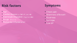
Transcript: Cardiology 11- 16- 2018 Case report HINT! INTRO TODAY'S SCHEDULE Ventricular Tachycardia 1. Basic Pathology 2. Risk factors & Symptoms 3. Diagnosis & Treatment 4. Case What? The Pathology Ventricular Arrhythmia Ventricular Arrythmia Types Types of VT Non-Reentrant VT * Ventricular pmc fire at higher rate, prevent SA node from firing ---> ventricular beats drive the HR * Due to: - Drugs (Amphetamine, cocaine etc) - Electrolyte imbalance - Myocardial ischemia in ventricle Reentrant VT (more common type) * Dead cells in myocardial tissue ---> Scar formation --> conduction signals go around scar --> Reentry Risk factors & signs Why? Risk & Symptoms Risk factors * Age * Cardiac disease (eg CMP, VD, post mi) * Electrolyte imbalance (Hyperkalemia) * Drugs (Cocaine) * Pericardial inflammation Symptoms * Chest pain * Shortness of breath * Dizziness * Syncope * Low BP Diagnosis * Serum electrolytes - K+ - Ca++ - Mg+ * ECG - Rate: >100 bpm - P-waves: Absent or Present- - T-waves: Large - QRS complex: Wide (>0,12 s) Diagnosis Treatment TREATMENT MEDICATION * Beta-blockers * Amiodarone * NDHP Ca++ ch. Blockers OTHER * Cardioversion * ICD Who? CASE Anamnesis: * Age: 51 * Sex: Female * Wide QRS * EF 35% --> 45% after medication * Hypokinetik inf. lat. & apical wall * Left ventricular edv 85 ml, 41mm * left ventricular systolic volume 47, 32mm * No pericardial fluid * MRI found scar Lab results * Troponin: Negative * Hc: Elevated * RBC: Elevated * WBC: Elevated * ALP: Elevated * Uric Acid: Elevated * K+: Slightly elevated

Cardiology Presentation

Transcript: What is a Cardiologist? -Focus on continuing care of patients. What does a day in a life of a cardiologist look like? First, they walk in their office. Then, they see their first patient of the day. After they meet their first patient, they ask about symptoms that the patient is exhibiting. Once the symptoms are known, the doctor starts to diagnose the patient by thinking how the symptoms relate to a disease. If needed, medical tests such as an echocardiogram or an angiogram are done in order to diagnose the patient. When the doctor arrives at a diagnosis, they tell the patient what it is and how to treat it. The doctor then advises the patient which treatment to take. Treatments include the usage of medication, a change in lifestyle, or even a medical procedure. The doctor will do this for about 4 or 5 more patients. The doctor might even take part in a procedure such as a radiofrequency ablation. First, the person completes undergraduate school (4 years) with an emphasis on science-related classes. Then, the pre-med student goes through medical school for four years. Then the doctor goes through a fellowship (2 years) with cardiologists to know the specifics of the field. Then the student becomes the cardiologist. What colleges offer these programs? Many colleges offer undergraduate programs in science, and even some offer pre-medicine programs. There also exist many medical schools in the United States, as well as the world. Once completing medical school, the medical school arrange a residency for you. A residency is completed at the hospital that the medical school chose for you. A cardiology fellowship is offered at many hopsitals. A radiofrequency ablation is used to treat SVT (Superventricular Tacchychardia) The success rate of a radiofrequency is over 90 percent. I personally have had this procedure, and I had no complications from it. Current Research being done in Cardiology... Dysfunctions in the diabetic heart. Risk factors for heart disease. The effects of kidney disease on the heart. or non-invasive (echocardiographies) A cardiologist normally sees patients with heart issues. "Radiofrequency Ablation". American Heart Association. 8.29.10 <http://www.strokeassociation.com/presenter.jhtml?identifier=4682>. Salamon, Maureen. "Do I Need to See a Cardiologist?". about.com. 8.29.10 <http://heartdisease.about.com/lw/Health-Medicine/Conditions-and-diseases/Do-I-Need-to-See-a-Cardiologist-.htm>. Cardiology is the field of medicine that studies and treats the heart. - A Cardiologist is a doctor that deals with heart problems. Then the post-medical student goes through a residency (3 years) at a hospital. What is the success rate of a radiofrequency ablation? -They also insert permanent and temporary cardiac pacemakers Cardiology Tim Klincewicz What kind of patients does a cardiologist see? Works Cited tests in order to diagnose patients. A normal cardiologist does not see patients under 18 years old. "Cardiologist". Schoolsintheusa.com. 8.29.10 <http://www.schoolsintheusa.com/careerprofiles_details.cfm?carid=508>. How many years of schooling does a Cardiologist need? What is Cardiology? Cardiology is the field of science that studies the heart and its associated functions. Cardiology is the field of science that studies the heart and its associated structures. Patients may range from deathly sick to not showing symptoms. "Cardiologist: Job Duties". degreedirectory.org. 8.29.10 <http://degreedirectory.org/articles/Cardiologist_Job_Duties_Occupational_Outlook_and_Education_Prerequisites.html>. -They use invasive (angiographies),

cardiology

Transcript: Thanks for your attention References ROC Curve r = -0.518 p = 0.003 MS에서 A fib까지 55mm p=0.04 p = 0.663 Results 65% (cc) image by anemoneprojectors on Flickr Statistical Analysis SPSS(version 19.0 SPSS Inc., Illinois, USA) Mean ± standard deviation 상관 분석은 피어슨 상관계수를 이용 P<0.05 인 경우 통계학적으로 유의한 것으로 판정 Limitation (cc) image by anemoneprojectors on Flickr Introduction p= 0.03 Mitral stenosis 80% Atrial fibrillation 각 그룹 별 LVEDD 관계 Introduction p= 0.161 Only MS 군에서 LVEDD/LADap와 DT의 관계 시간적으로 추적하지 못해 임상적 경과와 오차가 존재 다른 이완기능(EE’)이 누락된 환자가 많아서 더 좋은 이완기능 평가가 힘들었다 ROC Curve Limitation p = 0.02 승모판 협착증 환자에서 좌심실 이완기말 지름과 좌심방 전후 지름비와 그 합병증인 심방 세동과의 관계 p= 0.891 5+7= Conclusion Methods 한국 심초음파 학회: 임상심초음파학 제2판:2008,103P p= 0.936 (cc) image by anemoneprojectors on Flickr 민감도 김재원 sea8751626@hanmail.net p= 0.000 Summary AGENDA LLR와 DT의 관계 LLR과 TDI 비교 좌심방의 절대적인 크기였던 55mm 보다 본 연구에서 제시한 LVEDD/LADap 비율이 더 합리적이다. LVEDD/LADap 또한 좌심실의 기능과 연관이 있다. p = 0.03 p=0.726 Ratio of left ventricular end diastolic diameter to left atrial anteroposterior diameter is associated with atrial fibrillation in rheumatic mitral stenosis patients 특이도 p= 0.150 각 그룹 별 LVEF의 관계 MS with A fib 군에서 LVEDD/LADap와 LVEF의 관계 p= 0.000 Background 기존의 55mm 70% Subject 1. 환자 군 2003년 5월 부터 2012 5월까지 영남대학교 의료원 순환기 내과에 방문한 284명을 대상으로 함 2. 선정 기준 - 승모판 협착증을 갖고 있는 사람 승모판 협착증에 동반된 심방세동을 갖고 있는 사람 심방 세동만 갖고 있는 사람 연구 참가에 동의한 사람 3. 배제 기준 - 기저 심질환이 있는 환자: 심근경색, 협심증, 부정맥,고혈압 기저 내분비계 질환자: 당뇨병, 갑상선기능항진증 Participant characteristics p=0.000 Study design(1) 284명의 경 흉부 초음파를 시행한 환자를 대상으로 LVEDD,LADap,TDI,DT,LVEF등을 측정한 뒤 MS만 있는 군 MS와 심방 세동이 같이 존재하는 군 기저 질환 없이 심방 세동만 존재 하는 군 LVEDD/LADap Ratio 각 그룹별 LADap 관계 큰 집단을 이용한 민감도 예민도 연구가 필요 심방 세동과 좌심실의 관계에 대한 연구 환자를 선정하여 추적 관찰하는 연구 각 그룹 별 LLR 관계 Summary p= 0.067 r = -0.351 p= 0.011 1.Wolf PA,Daw TR, Thomas HE Jr, Kannel WB: Epidemiologic assessment of chronic atrial fibrillation and risk of stroke: the Framingham study.Neurology 1978;28(10):973-977 2.Roberts WC, Virmani R: Aschoff bodies at necropsy in valvular heart disease.Evidence from an analysis of 543 patients over 14 years of age that rheumatic heart disease, at least anatomically, is a disease of the mitral valve. Circulation 1978;57(4):803-807 3.Virmani R,Roberts WC:Aschoff bodies in operatively excised atrial appendages and in papillary muscles. Frequency and clinical significance.Circulation 1956;55(4):559-563 4.Pan M, Medina A, Suárez de Lezo J, et al:Factors determining late success after mitral balloon valvulotomy. Am J Cardiol 1993;71(13);1181-1185 5.Moreyra AE, Wilson AC, Deac R, et al:Factors associated with atrial fibrillation in patients with mitral stenosis: a cardiac catheterization study. Am heart J 1998;135(1):138-145 6.Aberg H.Atrial fibrillation. I. A study of atrial thrombosis and systemic embolism in a necropsy material. Acta Med Scand 1969;185(5):373-379 7.Stojnić BB, Radjen GS, Perisić NJ, Pavlović PB, Stosić JJ, Prcović M.:Pulmonary venous flow pattern studied by transoesophageal pulsed Doppler echocardiography in mitral stenosis in sinus rhythm: effect of atrial systole. Eur Heart J 1993;14(12):1597-1601 8.최윤식,이영우,제 2판 순환기학.일조각,2010,558P 9.심초음파 소위원회,박승우외: 한국인에서의 정상 심초음파 검사치의 확립에 관한 다기관연구 순환기 2000;30:373-82 10.Abhayaratna WP, Seward JB, Appleton CP, Douglas PS, Oh JK, Tajik AJ, Tsang TS.Left atrial size: physiologic determinants and clinical applications.J Am Coll Cardiol. 2006 Jun 20;47(12):2357-63. 11.Valocik G, Mitro P,Druzbacka L, Valocikova I. Left atrial volume as a predictor of heart function; Bratisl L Listy 2009;110(3)146-151 12.Jacob E. Møller, MD, PhD; Graham S. Hillis, MBChB, PhD.Left Atrial VolumeA Powerful Predictor of Survival After Acute Myocardial Infarction;Circulation 2003;107:2207-2212. 13.한국 심초음파 학회: 임상심초음파학 제2판:2008,103P. 14.Thamilarasan M, Klein AL. Factors relating to left atrial enlargement in atrial fibrillation: ‘chicken or the egg’ hypothesis. Am Heart J 1999; 137:381–383. p= 0.002 Braunwald’s heart disease 9th Left ventricle end diastole diameter / Left atrial anterior to posterior diameter ratio seems to be associate with the development of A fib in patients with MS (1.00± 0.14 vs 0.94± 0.12 p value 0.02) Left ventricle function also associate MS. Introduction Methods Results Conclusion http://www.cybermedk.com/MedicalInfo/Index.asp?Action=View&Gubun=topic&Mcheck=&Mform=&AccessKind=&AccessCode=&Idx=1007&Page=27 ROC Curve Conclusion 40% Introduction p= 0.078 p= 0.943 Future work 기존의 55mm The Routine use of warfarin in patients in sinus rhyrhm with LA enlargement (maximal dimension >5.5cm) with or without apontaneous echo contrast is more controversial. Harrison 18th edition Methods 김재원, 김웅, 박종선 영남대학교 의과대학 의학전문대학원 심장순환기 교실 Left ventricle end diastolic diameter/ Left atrium diameter anterior posterior Anticoagulation also may be considered for patients with severe MS and sinus rhythm when there is severe severe left atrial enlargement(55mm) 5+7= Background 승모판 협착증 p= 0.893 LVEDD/LADap Ratio Future work 65% 50% 20% 30% 기존의 55mm LVEDD/LADap Ratio 특이도

Now you can make any subject more engaging and memorable